Showing posts with label Lasik. Show all posts

What other types of refractive surgery are available?

Other types of refractive surgery are available and may be more appropriate than LASIK for certain individuals.

Advanced surface ablation: There are a variety of other techniques that utilize the excimer laser to reshape the cornea in much the same way as LASIK, but without the creation of a corneal flap. These are generically termed advanced surface ablation (ASA) and include photorefractive keratectomy (PRK), laser subepithelial keratomileusis (LASEK), and epipolis laser in situ keratomileusis (Epi-LASIK). All of these techniques involve first removing the most superficial corneal layer (epithelium) and then performing excimer laser ablation.

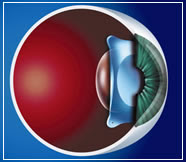
Phakic intraocular lenses: For patients with extreme myopia, LASIK and advanced surface ablation are not reasonable options. In these cases, a phakic intraocular lens may be used. This lens is implanted inside the eye and can effectively treat nearsightedness up to -20 diopters.

Conductive keratoplasty: Conductive keratoplasty (CK) is a technique that can be used for the temporary correction of hyperopia or presbyopia. CK involves using radiofrequency waves in the peripheral cornea to cause peripheral corneal shrinkage and central steepening. This procedure is very safe, but its effect is often not long-lasting, and regression is common after a few years.

Intracorneal ring segments: Intacs (Addition Technology, Inc.) are approved for the correction of low myopia and for patients with keratoconus in the U.S. Intacs are micro-thin plastic segments that are implanted into the peripheral cornea in order to flatten the cornea centrally. Once implanted, the rings generally cannot be felt by the patient. These rings can be removed, and their effect is usually completely reversible. They are only able to correct up to -3 diopters of myopia, and visual recovery is generally slower and less predictable than LASIK.

LASIK Eye Surgery

During our youth, the natural lens has the ability to change shape and power. This allows us to focus on close objects through a process of lens power change called accommodation. As we age, the natural lens becomes stiffer and loses the ability to change shape. This is termed presbyopia, which is the loss of accommodation, and the need for reading glasses, bifocals, or other visual aids to facilitate near work. LASIK cannot directly "fix" accommodation, but there are a variety of strategies that can be successful, including blended vision or monovision, in which one eye is corrected for better distance vision and one eye is corrected for better near vision.

effective for thin-flap LASIK

effective for thin-flap LASIKA single-use microkeratome with disposable components is safe and effective for creating flaps in thin-flap LASIK and is a viable alternative to femtosecond lasers for this purpose, Gustavo Tamayo, MD, told ophthalmologists gathered at the recent annual meeting of the American Society of Cataract and Refractive Surgery (ASCRS).

Thin-flap LASIK, also called sub-Bowman's keratomileusis (SBK), involves creating a thinner flap than flaps traditionally produced in LASIK surgery.

A thinner flap may reduce the risk of dry eyes after LASIK. It also may preserve greater corneal strength, thereby reducing the risk of corneal ectasia after the procedure.

Using a microkeratome in

thin-flap LASIK also may lower LASIK costs, as the equipment costs for these mechanical instruments are significantly less for LASIK surgeons than costs associated with femtosecond lasers.


Single-use microkeratome: Study design and methods ( nowGoogle.com adalah Multiple Search Engine Popular)

Dr. Tamayo conducted a study at Bogota Laser Refractive Institute (Bogota, Columbia) to compare the outcomes of 40 eyes undergoing thin-flap LASIK with corneal flaps created with the Moria One Use-Plus single-use microkeratome or a femtosecond laser.

In all cases, the intended flap thickness was 100 microns.

Prior to surgery, the mean amount of myopia in the microkeratome group was -1.50 D. In the femtosecond laser group, it was -2.25 D.

Flap thickness was measured with an ultrasonic device during surgery immediately after the flaps were created.


Single-use microkeratome: Study results

Analysis of data gathered in the study revealed:

* Mean flap thickness was very similar in the two groups: 101 microns in the microkeratome group (range: 85 to 108 microns) and 104 microns in the femtosecond laser group (range: 82 to 110 microns).

* Corneas in the microkeratome group showed slightly more decrease in biomechanical stability factors than corneas in the femtosecond laser group.

(Note: a 30-micron range is equivalent to 0.3 millimeter.)

Dr. Tamayo said other larger studies also have shown the microkeratome is capable of producing thin flaps with excellent reproducibility of flap dimensions.

In addition, these studies have demonstrated that thin flaps created with the Moria microkeratome are nearly planar (of equal thickness throughout) and the underlying corneal bed after flap creation with the microkeratome is smoother than the underlying corneal bed after flap creation with a 60-kHz femtosecond laser.


Single-use microkeratome: Conclusions

Dr. Tamayo concluded that the Moria One Use-Plus microkeratome with disposable, single-use components is safe, effective and reliable for creating flaps in thin-flap LASIK, and it provides excellent outcomes at a lower cost than femtosecond lasers. nowGoogle.com

LASIK - Laser Eye Surgery

The refractive state of your eye can be improved and you can decrease the need for wearing glasses through Refractive Eye Surgery. The most common method of surgery followed today is Laser surgery, where lasers are used to reshape the cornea. Laser surgery is the greatest achievement in ophthalmology. Lasers are usually used to treat non refractive conditions.

What is LASIK?

LASIK is one of the most popular types of laser eye surgery. LASIK stands for Laser-Assisted In Situ Keratomileusis. This procedure can permanently reshape the cornea, which is the clear covering of the front of the eye, and change its focus power using an excimer laser. Millions of patients have been treated using LASIK, using of a wide variety of different lasers. This procedure can be used to correct myopia, hyperopia and astigmatism.

Prior to the surgery:

Before you undergo LASIK surgery you

will have to get your eyes examined by a doctor to see if you are a good candidate. If you wear contact lenses, you will have to stop using them for 1 to 4 weeks before the surgery and switch to wearing your glasses, to allow the cornea to return to its original shape, or else this can have negative consequences like inaccurate measurements and a poor surgical plan; resulting in poor vision after surgery. These measurements would decide how much corneal tissue is to be removed, and have to be repeated a week after your first evaluation as well as before surgery, to see that the measurements have not changed.

It is important that you tell your doctor all about your past and present medical eye conditions, and also the medications you are taking or if you are allergic to any medicines. Then on the day of the surgery you will be given a numbing drop so that you do not feel pain, although some patients do experience some discomfort. The area around your eye will be cleaned and a lid speculum will be used, which is an instrument to hold your eyelids open. A suction ring will be placed on your eye and suction will be created by applying very high pressure to the cornea. As a result of this, your vision will dim and the pressure may cause some discomfort.

During the surgery:

A small device called a microkeratome will be attached to the suction ring; the blade of which will be used to cut a thin flap on your cornea. The flap is created, exposing cornea tissue. After this, both the microkeratome and the suction ring will be removed. The doctor will now lift the flap and fold it back on its hinge, exposing a layer of tissue called the stroma - the middle section of the cornea.

You will be made to focus on a particular spot and the doctor will begin the laser to remove the corneal tissue, according to the measurements taken at the initial examination. The flap is then put back into position and left to heal.

After the surgery:

As no stitches are required, you will just have to wear an eye patch as protection to prevent rubbing, getting poked, or any kind of pressure that might disturb the healing process. It may take up to three to six months for your vision to stabilize after surgery.

It is important that you know all about Laser Surgery before you go through with it - the benefits as well as the side effects if there are any. Consult with your doctor, as only he will be able to tell you if this surgery will be good for you. Once you have all the information needed, you can make the decision whether or not you wish to go ahead with the surgery, and give yourself the opportunity to see the world in a whole new way!
by : Paul MacIver